
In short. Wood's lamp examination, the conditions that mimic vitiligo, and why a dermatologist has to confirm the diagnosis before treatment starts.
Key points
- Treating the wrong diagnosis wastes months and can cause harm.
- A potent topical steroid applied to a fungal infection makes it worse, and phototherapy given for a condition that is not vitiligo exposes the person to ultraviolet for no reason.
Vitiligo is usually diagnosed at the bedside, and the examination is more informative than any blood test. A dermatologist looks at where the patches sit, how sharp their borders are, whether the pigment loss is complete or partial, and whether the pattern is symmetrical. The distribution carries a great deal of information: vitiligo has a strong preference for the area around the eyes and mouth, the fingers and wrists, the elbows and knees, the shins, and the body folds.
The Wood's lamp
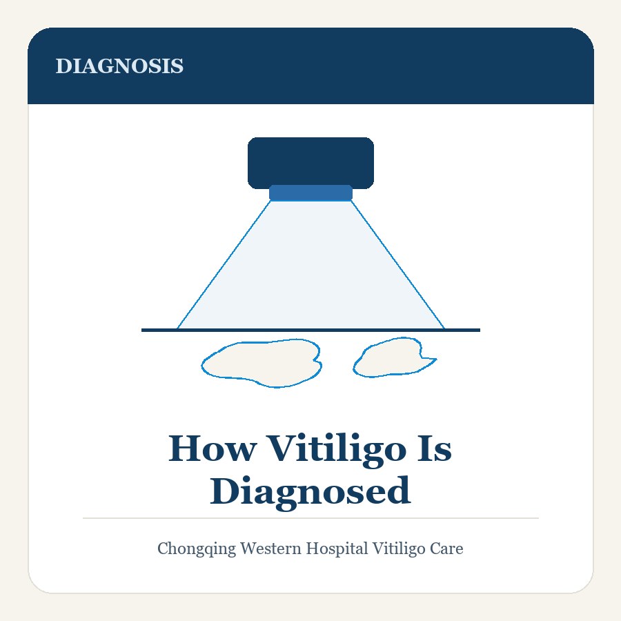
A Wood's lamp is a filtered ultraviolet light that makes pigment loss far easier to see. Under it, depigmented skin glows a bright blue-white against the surrounding skin, and partial pigment loss that is easy to miss in ordinary light becomes obvious. It lets the clinician map how much skin is involved, find patches the person had not noticed, and distinguish complete depigmentation from the partial lightening seen in other conditions. It is painless and takes a few minutes.
Occasionally a small skin sample is taken, usually when the diagnosis is genuinely unclear or when another cause of pigment loss is possible. Most people do not need one.
What else looks like vitiligo
Several conditions produce pale patches, and they are treated very differently.
Pityriasis versicolor is a common fungal condition that produces pale, slightly scaly patches on the trunk and shoulders, often more noticeable after sun exposure darkens the surrounding skin. It is not vitiligo, it is not autoimmune, and it responds to antifungal treatment. The fine scale and the distribution are the clues.
Post-inflammatory hypopigmentation follows injury, eczema, psoriasis, or an infection, and the pale area matches where the inflammation was. It usually improves over months as the skin recovers, unlike vitiligo, which does not repigment on its own.
Other possibilities include a mole that has lost its pigment, a birthmark such as nevus depigmentosus, and the pale patches of tuberous sclerosis. Some medicines and chemicals can also lighten skin. None of these is managed in the same way as vitiligo.
Why confirmation matters
Treating the wrong diagnosis wastes months and can cause harm. A potent topical steroid applied to a fungal infection makes it worse, and phototherapy given for a condition that is not vitiligo exposes the person to ultraviolet for no reason. Confirmation comes first.
Arrange a dermatology assessment if
- Pale or white patches are appearing, spreading, or changing shape.
- A pale patch is scaly, itchy, or has lost sensation.
- A mole has become paler, or a patch has developed a raised, firm, or irregular area within it.
Frequently asked questions
How is vitiligo diagnosed, and is a blood test needed?
It is usually diagnosed at the bedside. The dermatologist looks at where the patches sit, how sharp their borders are, whether pigment loss is complete or partial, and whether the pattern is symmetrical. The examination is more informative than any blood test.
What does a Wood’s lamp add to the examination?
It is a filtered ultraviolet light. Depigmented skin glows bright blue-white against the surrounding skin, so partial pigment loss that is easy to miss in ordinary light becomes obvious. It also lets the clinician map how much skin is involved and find patches the person had not noticed. It is painless and takes a few minutes.
What conditions can be mistaken for vitiligo?
Pityriasis versicolor, a fungal condition with pale, slightly scaly patches on the trunk and shoulders, is one. Post-inflammatory hypopigmentation follows injury, eczema or psoriasis and usually improves over months. Others include a mole that has lost its pigment, a birthmark, and the pale patches of tuberous sclerosis.
Risks & Limitations
- Repigmentation is slow, measured in months to years, and is often partial rather than complete.
- Vitiligo is unpredictable: treatment can control it, but nothing guarantees that it will stop spreading.
- Treatment usually needs to continue to hold the result, and relapse is common after it is stopped.
- Prolonged topical steroid use can thin the skin, cause visible blood vessels and stretch marks, and trigger outbreaks of small spots around the hair follicles.
- Phototherapy can burn, and the ultraviolet dose accumulates over a lifetime, raising the risk of skin ageing and skin cancer.
- The face and neck respond best; the hands, feet and lips are the hardest areas, where pigment may never return.
- No treatment works for everyone, and the response of an individual patch cannot be predicted with certainty.
When to Seek Care
- Depigmentation is spreading rapidly, or new patches are appearing over weeks rather than months.
- Depigmentation involves the eyelids or the skin around the eyes.
- New symptoms suggest a thyroid or other autoimmune condition — persistent fatigue, weight change, feeling cold, a neck swelling, or excessive thirst.
- Blistering, burning or severe pain after a phototherapy session — stop treatment and seek advice.
- Eye pain, redness, light sensitivity or blurred vision, which need prompt assessment.
References & Further Reading
- American Academy of Dermatology (AAD) — public and clinical guidance on vitiligo
- Vitiligo Research Foundation — patient and clinician information resources
- Cochrane Library — systematic reviews of vitiligo treatment
- Chinese Medical Association, Dermatology Branch — clinical guidance
This page is patient education, not medical advice, and it does not cite individual studies. Discuss your own case with a qualified clinician. To arrange an assessment at Chongqing West District Hospital, see Contact & Consultation.